Long Bone Diagram Endosteum - 6 3 Bone Structure Anatomy Physiology / Spongy bone is composed of trabeculae that contain the osteocytes.. A long bone has a shaft and 2 ends. The diaphysis is the hollow, tubular shaft that runs between the proximal and distal ends of the bone. Osteocytes & chondrocytes live in small spaces in the matrix called ____.bone anatomy long diagram human marrow cell osteoporosis spongy matrix medical vector joint structure system body compact disease endosteum illustration periosteum anatomical biology blood calcium care cartilage diaphysis educational epiphysis femur health healthy. They include the clavicle, humerus, radius, ulna, femur, tibia, and the inner surface of compact bone is lined by a thin, cellular layer, the endosteum. The diaphysis and the epiphysis (figure 6.3.1).
The medullary cavity, the hollow spaces in the trabecular (spongy) bone, haversian (osteonic) and volkmann's (perforating) canals in the cortical (compact) bone of the long bones, such as humerus and femur, flat bones, such as ribs 5 and pelvic bones 6, and sesamoid bones, such as patella 10. This membrane is found in the diaphysis, or shaft, of long bones. Long bones are those in which the length exceeds the breadth and thickness. Endosteum is a thin, soft, connective tissue, lining the cavity of long bones like humerus and femur. Red marrow fills the spaces in some bones.
Bone Structure Anatomy And Physiology from s3.amazonaws.com A long bone has two main regions: It is a thin covering that surrounds the medullary cavity. Long, short, flat, irregular and sesamoid. Long bone diagram endosteum : Long bones are those in which the length exceeds the breadth and thickness. Definition and functions the endosteum is a structure in the middle of bone tissue and bone marrow. Long bones include the humerus (upper arm), radius (forearm), ulna (forearm), femur (thigh), fibula (thin bone of the lower leg), tibia (shin bone), phalanges (digital bones in the hands and feet), metacarpals (long bones within the hand), and metatarsals (long bones within. A long bone has two parts:
This endosteal surface is usually resorbed during long periods of malnutrition, resulting in less cortical thickness.
1 endosteum has cells known as endosteal. Long bones are found in the arms (humerus, ulna, radius) and legs (femur, tibia, fibula), as well as in the fingers (metacarpals, phalanges) and toes (metatarsals, phalanges). The diaphysis and the epiphysis (figure 6.3.1). Long bones are one of the five bone types that are classified by shape. A diagram of the anatomy of a bone, showing the endosteum. The endosteum (plural endostea) is a thin vascular membrane of connective tissue that lines the inner surface of the bony tissue that forms the medullary cavity of long bones. The outer and inner regions contain layers of lamellar bone that run circumferentially around the entire bone. Inside the diaphysis is the medullary cavity, which is filled with yellow bone marrow in an adult. These include medullary cavity and medullary membrane. The outer surface of a bone is lined by a thin layer of connective tissue that is very similar in. Diagramme schnell und einfach erstellen. The endosteum is lined with a single thin layer of bone lining cells (mature osteoblasts) and osteoblasts which form a membrane over endocortical and trabecular bone surfaces to enclose the bone marrow. A long bone has a shaft and 2 ends.
The endosteum can be seen in the t.s. The medullary cavity, the hollow spaces in the trabecular (spongy) bone, haversian (osteonic) and volkmann's (perforating) canals in the cortical (compact) bone of the long bones, such as humerus and femur, flat bones, such as ribs 5 and pelvic bones 6, and sesamoid bones, such as patella 10. Endosteum is located in bones such as femur, humerus, hip bone, thoracic rib bones and sesamoid bones like patella. 30 osteoclasts can also be present in the endosteum in regions of active bone resorption. A long bone has diaphyseal bone is organized to create the best balance between weight and structural strength.
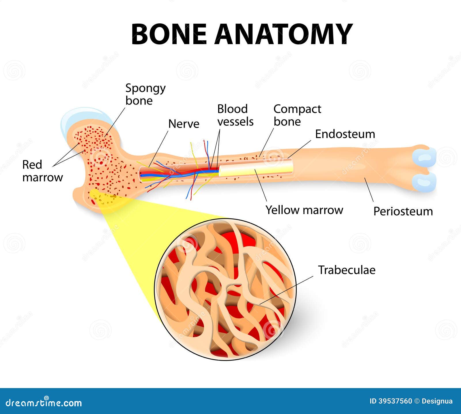
Bone Anatomy Stock Vector Illustration Of Marrow Body 39537560 from thumbs.dreamstime.com The periosteum forms the outer surface of bone, and the endosteum lines the medullary cavity. This is covered by a membrane of connective tissue called the periosteum.beneath the cortical bone layer is a layer of spongy cancellous bone.inside this is the medullary cavity which has an inner core of bone marrow, it contains nutrients and help in formation of cells, made up of yellow marrow in. Endosteum is a thin, soft, connective tissue, lining the cavity of long bones like humerus and femur. The medullary cavity, the hollow spaces in the trabecular (spongy) bone, haversian (osteonic) and volkmann's (perforating) canals in the cortical (compact) bone of the long bones, such as humerus and femur, flat bones, such as ribs 5 and pelvic bones 6, and sesamoid bones, such as patella 10. The endosteum can be seen in the t.s. Osteocytes & chondrocytes live in small spaces in the matrix called ____.bone anatomy long diagram human marrow cell osteoporosis spongy matrix medical vector joint structure system body compact disease endosteum illustration periosteum anatomical biology blood calcium care cartilage diaphysis educational epiphysis femur health healthy. Red marrow fills the spaces in some bones. Start studying (a&p 1) parts of the long bone.
Spongy bone is composed of trabeculae that contain the osteocytes.
This layer of membrane envelopes the spongy tissue, the medullary cavity and the internal lining of the bone's cavity and the haversian canal of the marrow. A long bone has a shaft and 2 ends. The diaphysis is the hollow, tubular shaft that runs between the proximal and distal ends of the bone. Endosteum is a soft, thin connective tissue that lines the inner cavity of long bones. 30 osteoclasts can also be present in the endosteum in regions of active bone resorption. This endosteal surface is usually resorbed during long periods of malnutrition, resulting in less cortical thickness. Learn vocabulary, terms, and more with flashcards, games, and other study tools. The medullary cavity, the hollow spaces in the trabecular (spongy) bone, haversian (osteonic) and volkmann's (perforating) canals in the cortical (compact) bone of the long bones, such as humerus and femur, flat bones, such as ribs 5 and pelvic bones 6, and sesamoid bones, such as patella 10. Lumbar spine, femoral neck, distal radius) to determine bone. The endosteum is lined with a single thin layer of bone lining cells (mature osteoblasts) and osteoblasts which form a membrane over endocortical and trabecular bone surfaces to enclose the bone marrow. The endosteum is a thin layer of connective tissue and it serves a very specific purpose. Start studying (a&p 1) parts of the long bone. Inside the diaphysis is the medullary cavity, which is filled with yellow bone marrow in an adult.
30 osteoclasts can also be present in the endosteum in regions of active bone resorption. The periosteum forms the outer surface of bone, and the endosteum lines the medullary cavity. Long, short, flat, irregular and sesamoid. The endosteum appears at the interface between the cortical bone and the medullary cavity in long bones and with pathology, may appear scalloped (see endosteal scalloping). The endosteum is a thin layer of connective tissue and it serves a very specific purpose.
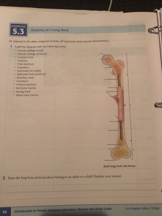
Solved Exercise 5 3 Anatomy Of A Long Bone As Opposed To Chegg Com from media.cheggcdn.com The outer and inner regions contain layers of lamellar bone that run circumferentially around the entire bone. The thigh bone (femur) is a long bone. The endosteum can be seen in the t.s. The endosteum is located on the internal surface of the bone, being the membranous layer that covers the medullary cavity, the bony trabeculae (spongy part of the bone), the haversian canals and internal walls of the compact long bones. Periosteum, endosteum, bone marrow and trabeculae. A long bone is a bone that has greater length than width. The diaphysis is the hollow, tubular shaft that runs between the proximal and distal ends of the bone. The periosteum forms the outer surface of bone, and the endosteum lines the medullary cavity.
30 osteoclasts can also be present in the endosteum in regions of active bone resorption.
Learn vocabulary, terms, and more with flashcards, games, and other study tools. The diaphysis is the hollow, tubular shaft that runs between the proximal and distal ends of the bone. Long bones include the humerus (upper arm), radius (forearm), ulna (forearm), femur (thigh), fibula (thin bone of the lower leg), tibia (shin bone), phalanges (digital bones in the hands and feet), metacarpals (long bones within the hand), and metatarsals (long bones within. The thigh bone (femur) is a long bone. Figure 6.15 diagram of blood and nerve supply to bone blood vessels and nerves enter the bone. It is a thin covering that surrounds the medullary cavity. Lumbar spine, femoral neck, distal radius) to determine bone. The endosteum is lined with a single thin layer of bone lining cells (mature osteoblasts) and osteoblasts which form a membrane over endocortical and trabecular bone surfaces to. The main type of bone cell is the osteocyte (bone cell, shown as purple in the diagram). A long bone is one that is cylindrical in shape, being longer than it is wide.keep in mind, however, that the term describes the shape of a bone, not its size. Osteocytes & chondrocytes live in small spaces in the matrix called ____.bone anatomy long diagram human marrow cell osteoporosis spongy matrix medical vector joint structure system body compact disease endosteum illustration periosteum anatomical biology blood calcium care cartilage diaphysis educational epiphysis femur health healthy. Diagram of blood and nerve supply to bone. Endosteum is found on all internal surfaces of bones:
1 endosteum has cells known as endosteal long bone diagram. The endosteum (plural endostea) is a thin vascular membrane of connective tissue that lines the inner surface of the bony tissue that forms the medullary cavity of long bones.


0 Komentar